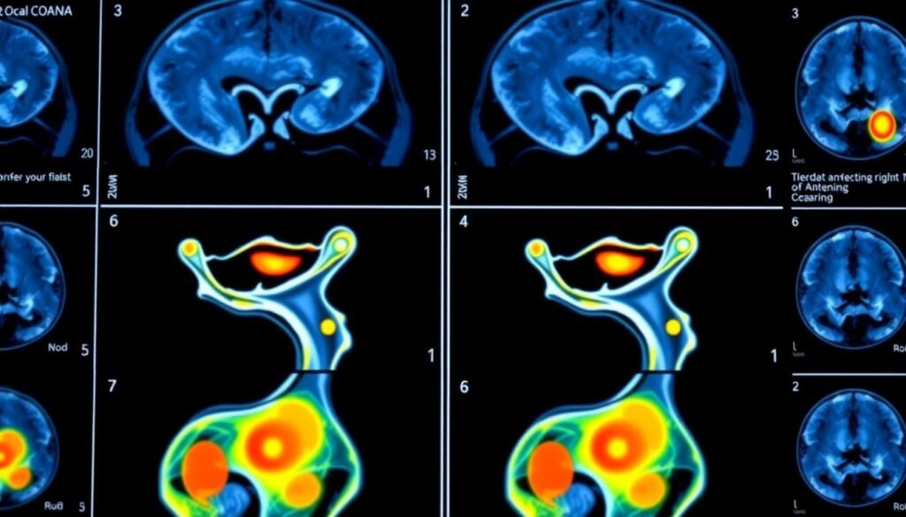
What role does radiology play in figuring out cervical cancer’s stage? How do imaging techniques affect treatment plans? Cervical cancer staging through radiology is key for diagnosis and treatment. It uses different imaging methods to see how far the disease has spread.
Techniques like MRI and CT scans give doctors important details. They show the tumor’s size, where it is, and if it has spread. This info is crucial for figuring out the cancer’s stage. Knowing radiology’s role helps patients understand their diagnosis and treatment better.
Table of Contents
Key Takeaways
- Radiology plays a critical role in cervical cancer staging
- Cervical cancer imaging techniques, such as MRI and CT scans, are used to determine the stage of the disease
- Accurate staging is essential for informing treatment decisions
- Cervical cancer staging radiology helps doctors understand the severity of the disease
- Imaging techniques provide valuable information about the tumor’s size, location, and spread
Understanding the Basics of Cervical Cancer Staging
Cervical cancer diagnostic imaging is key in finding out how far the disease has spread. Knowing the extent of the disease is crucial for creating effective treatment plans. Imaging tests help see the tumor’s size, where it is, and if it has spread.
When it comes to cancer staging, accuracy is key. Doctors look at the tumor’s size, location, and if it has spread. This helps them decide the best treatment and what to expect. For cervical cancer, radiological staging is a big part of figuring out the diagnosis.
What is Cancer Staging?
Cancer staging is complex. It looks at the tumor’s size, location, and spread. This info helps doctors plan the best treatment. For cervical cancer, radiological staging of cervical cancer is very important.
The Importance of Accurate Staging
Getting the staging right is key for good treatment and outcomes. For cervical cancer, cervical cancer diagnostic imaging is vital. It helps doctors plan the best treatment and improve patient results.
FIGO Staging System Overview
The FIGO staging system is used a lot for cervical cancer. It looks at the tumor’s size, location, and spread. This system helps doctors figure out the disease stage and plan treatment. Using radiological staging of cervical cancer with the FIGO system helps improve treatment plans and patient outcomes.
The Critical Role of Radiological Assessment in Cervical Cancer
Radiologic evaluation is key in finding out how far cervical cancer has spread. It uses different imaging methods to check the cancer’s stage. This helps doctors plan the best treatment.
Imaging is vital for understanding the cancer’s extent. It looks at the main tumor, lymph nodes, and if the cancer has spread. This info helps doctors create a good treatment plan.
Imaging methods are important for getting accurate cancer information. Some key things in radiologic assessment are:
- Evaluating the size and location of the primary tumor
- Assessing lymph node involvement
- Detecting distant metastases
Accurate staging is crucial for choosing the right treatment. Radiologic evaluation helps doctors decide between surgery, radiation, or other treatments. With the latest imaging, patients get care that fits their needs.
Key Imaging Modalities for Cervical Cancer Staging Radiology
Several imaging modalities are key in cervical cancer staging radiology. These tools help accurately assess the disease. A mix of these modalities is often used to understand the cancer’s extent and spread.
Magnetic Resonance Imaging (MRI)
MRI is great for finding cervical cancer early. It shows detailed images of the tumor and nearby tissues. This helps doctors see how far the cancer has spread.
Computed Tomography (CT)
CT scans are used to stage cervical cancer. They help see if the cancer has spread to distant organs. They show the tumor’s size, location, and how it relates to nearby structures.
PET-CT Scanning and Ultrasound Applications
PET-CT scans and ultrasound add more info on cervical cancer. They show the disease’s metabolic activity and spread to lymph nodes and other areas.
By following cervical cancer radiology guidelines, radiologists can give accurate assessments. This helps guide treatment and improves patient outcomes.
| Imaging Modality | Advantages | Limitations |
|---|---|---|
| MRI | High sensitivity, detailed images | Cost, availability |
| CT | Wide availability, fast imaging | Radiation exposure, lower sensitivity |
| PET-CT | Metabolic activity information, whole-body imaging | High cost, limited availability |
| Ultrasound | Non-invasive, low cost | Limited depth penetration, operator-dependent |
MRI: The Gold Standard in Cervical Cancer Assessment
Magnetic Resonance Imaging (MRI) is the top choice for advanced imaging in cervical cancer staging. It’s very good at finding the main tumor and checking if cancer has spread to lymph nodes. This helps doctors decide the best treatment.
There are many reasons MRI is better than other imaging methods. It shows the tumor and nearby tissues clearly. This is key for knowing how to treat the cancer, like surgery or radiation.

In advanced imaging in cervical cancer staging, MRI’s role in checking lymph nodes is huge. It tells doctors if cancer has reached the lymph nodes. This info helps plan treatments that might include special therapies or stronger treatments.
Using MRI in cervical cancer checks has a big effect on patient results. It gives doctors the exact details they need to choose the right treatment. This leads to better care and higher survival rates for patients.
Advanced Imaging Techniques and Their Impact
New imaging technologies have greatly improved how we find and stage cervical cancer. Techniques like diffusion-weighted imaging and dynamic contrast enhancement have made a big difference. They give doctors a better look at the tumor, helping them make better choices.
Diffusion-weighted imaging lets doctors see how dense the tumor is. This is key for figuring out the cancer’s stage. Dynamic contrast enhancement also helps by showing the tumor’s blood flow. This info is vital for staging the cancer.
Artificial intelligence is also changing cervical cancer imaging. AI can look through lots of data to find patterns and problems. This could lead to even more accurate diagnoses and better care for patients.
Key Benefits of Advanced Imaging Techniques
- Improved accuracy in cervical cancer staging radiology
- Enhanced tumor characterization
- More informed treatment decisions
- Potential for improved patient outcomes
As research keeps moving forward, we’ll likely see even more advanced imaging tools. These will help make cervical cancer diagnosis and treatment even better.
Interpreting Radiological Findings in Cervical Cancer
Getting radiological findings right is key in cervical cancer diagnostic imaging. It shows how far the disease has spread. This info is crucial for making a good treatment plan. Radiologists are important here, as they look at images from MRI and CT scans.
They check the tumor’s size, where it is, and if it has spread. This helps in planning the treatment.
In radiological staging of cervical cancer, being consistent and accurate is vital. Radiologists often work together. They review and talk about the findings to make sure they have all the info.
This teamwork helps lower mistakes and makes the staging more reliable.
Some good ways to interpret radiological findings include:
- Using standardized reporting templates to ensure consistency
- Correlating imaging findings with clinical and pathological data
- Staying up-to-date with the latest imaging technologies and techniques

By following these tips and using the newest in cervical cancer diagnostic imaging and radiological staging of cervical cancer, doctors can get better at staging. This leads to better care for cervical cancer patients.
Limitations and Challenges in Radiological Staging
The radiologic evaluation of cervical cancer is key in figuring out how far the disease has spread. Yet, there are big challenges in this area. The main issue is the technical limits of imaging tools for cervical cancer staging, which can cause wrong assessments.
Things like motion artifacts and metal artifacts can mess up the accuracy. These can be lessened by using the latest imaging methods and rules. Also, issues like obesity and claustrophobia in patients can affect how well images are seen and understood.
To tackle these problems, it’s vital to mix different imaging methods for cervical cancer staging, like MRI, CT, and PET-CT. This way, doctors can get a clearer picture of the disease and give accurate stages. The American College of Radiology says, “Using many imaging methods can make staging more accurate and help patients get better care.”
- Use advanced imaging techniques like diffusion-weighted imaging and dynamic contrast enhancement.
- Apply artificial intelligence algorithms to help with image reading.
- Keep radiologists up-to-date with the newest imaging tools and methods.
By facing and solving the issues with radiological staging, we can aim to make the radiologic evaluation of cervical cancer more precise. This will help us give better care to our patients.
Future Developments in Cervical Cancer Imaging
The field of cervical cancer imaging is always changing. New technologies and techniques are being developed to help diagnose and stage the disease better. Recent cervical cancer radiology guidelines highlight the growing importance of advanced imaging like MRI and PET in the radiological assessment of cervical cancer.
Research and development in cervical cancer imaging are focusing on several key areas:
- Emerging technologies like artificial intelligence (AI) and machine learning (ML) are being explored to enhance image analysis and interpretation.
- Advances in imaging modalities, such as MRI and PET, are providing clearer and more detailed images of tumors.
- New contrast agents are being developed to improve the visibility of tumors and surrounding tissues.
These advancements are set to significantly impact cervical cancer diagnosis and staging. They are expected to lead to better patient outcomes. It’s crucial to keep up with the latest cervical cancer radiology guidelines and radiological assessment of cervical cancer to provide top-notch care for patients.

By integrating these new technologies and techniques into clinical practice, healthcare providers can enhance diagnosis accuracy and develop more effective treatment plans. This will result in better patient outcomes and improved quality of life for those with cervical cancer.
| Technology | Description |
|---|---|
| MRI | High-resolution imaging modality used to visualize tumors and surrounding tissues |
| PET | Imaging modality used to visualize metabolic activity of tumors |
| AI/ML | Technologies used to improve image analysis and interpretation |
Best Practices for Optimal Imaging Results
Getting the best results from advanced imaging in cervical cancer staging is key. This means preparing patients right, choosing the right imaging tools, and understanding cervical cancer radiology findings well.
For top-notch imaging, doctors and radiologists must keep up with new guidelines and tech. They need ongoing learning to give accurate and dependable cervical cancer radiology findings.
Here are some tips for better imaging results:
- Choose the best imaging method for each patient
- Make sure patients are well-prepared and positioned right
- Use advanced techniques like diffusion-weighted imaging and dynamic contrast enhancement
By sticking to these best practices and keeping up with new advanced imaging in cervical cancer staging, doctors can give accurate cervical cancer radiology findings. This helps improve patient care.
| Imaging Modality | Description |
|---|---|
| Magnetic Resonance Imaging (MRI) | Provides detailed images of the cervix and surrounding tissues |
| Computed Tomography (CT) | Helps identify the extent of cancer spread and lymph node involvement |
Conclusion: The Future of Radiological Staging in Cervical Cancer
As we wrap up our look at cervical cancer staging radiology, it’s clear that new imaging methods have changed how doctors diagnose and treat this disease. MRI, CT, and PET-CT scans are now key in figuring out how far cancer has spread. This makes treatment planning much better.
The future of cervical cancer imaging techniques looks very promising. New tech like diffusion-weighted imaging and dynamic contrast enhancement will give even more detailed views. Also, artificial intelligence will help doctors make quicker and better decisions. Working together, experts will keep making these advances.
Using radiology to stage cancer means doctors can give patients the best treatment. This leads to better health and life quality. As cervical cancer staging radiology keeps getting better, we’re looking forward to even more precise care for patients.
FAQ
What is the role of radiology in cervical cancer staging?
Radiology is key in accurately staging cervical cancer. Imaging like MRI, CT, and PET-CT helps check the tumor size, lymph nodes, and distant spread. These steps are vital for choosing the right treatment and predicting outcomes.
What are the key imaging modalities used for cervical cancer staging?
The main imaging tools for cervical cancer staging are:
– Magnetic Resonance Imaging (MRI): The top choice for looking at the tumor and its spread.
– Computed Tomography (CT): Good for spotting lymph nodes and distant cancer.
– Positron Emission Tomography (PET-CT): Helps find hidden cancer and plan treatment.
– Ultrasound: Useful for seeing how deep the tumor is and checking lymph nodes.
Why is MRI considered the gold standard for cervical cancer assessment?
MRI is the best for cervical cancer because it’s very good at finding tumors and seeing how they spread. Its clear images and ability to view from different angles make it the top choice for accurate staging and treatment planning.
What are some advanced imaging techniques used in cervical cancer staging?
New imaging methods for cervical cancer include:
– Diffusion-Weighted Imaging (DWI): Shows tumor details like cell density.
– Dynamic Contrast Enhancement (DCE): Looks at tumor blood flow and growth.
– Artificial Intelligence (AI) Integration: Uses AI to help read and analyze images, aiming for better accuracy.
What are some of the limitations and challenges in radiological staging of cervical cancer?
Challenges in cervical cancer imaging include:
– Limitations of imaging tech, like resolution and contrast.
– Patient factors like obesity can impact image quality.
– Difficulty in telling benign from malignant findings.
– Need for radiologists to keep up with new imaging tech.
What are the best practices for optimal imaging results in cervical cancer staging?
For the best imaging results, follow these steps:
– Proper patient prep, like full bladder and bowel prep.
– Choose the right imaging modality based on the case.
– Stick to imaging protocols and guidelines for quality studies.
– Work together with doctors and radiologists for a complete care plan.


